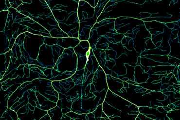
Our nervous system is composed of billions of neurons that speak to each other through their axons and dendrites. When the human brain develops, these structures branch out in a beautifully intricate, yet poorly understood, way that allows nerve cells to form connections and send messages throughout the body. And now, Yale researchers have discovered the molecular mechanism behind the growth of this complex system. Their findings are published in Science Advances.
“Neurons are highly branched cells, and they’re like this because each neuron makes a connection with thousands of other neurons,” says Joe Howard, PhD, Eugene Higgins Professor of Molecular Biophysics and Biochemistry and professor of physics, and senior researcher of the study. “We’re working on this branching process—how do branches form and grow? That is what’s underlying the whole way the nervous system works.”
The team studied neuronal growth in fruit flies as they matured from embryos into larvae. To visualize this process, they tagged neurons with fluorescent markers and imaged them on a spinning disk microscope. Because neurons reside just under the cuticle [outermost layer], the researchers were able to observe this process in real time in live larvae. After imaging the neurons at different stages of development, the team was able to create time-lapse movies of the growth.
This story was excerpted from the Yale School of Medicine News story of August 2, 2022. Click below for the full story and for a link to the article in Science Advances.